


Last month you could learn about the technological capabilities of the Thermo Phenom desktop SEM in our blog on desktop SEM “SEM in the reach of every lab.” In response, we received quite a few questions related to the possibilities that the EDS (energy-dispersive x-ray spectroscopy) module has to offer as an analytical complement to the standard imaging capabilities of the SEM. Often the main topics of interest then are resolution, quantification, peak separation and the software capabilities for surface scanning and line scanning, among others.
In this follow-up article, we will take a closer look at the capabilities that desktop SEM has to offer when we start combining SEM image analysis with chemical analysis via EDS.
Enhancing SEM applications by using EDS
EDS provides information on the composition of materials by analyzing the characteristic X-rays generated by the interaction of the sample with the electron beam. The high brightness of the CeB6 and FEG sources in a Thermo Scientific Phenom SEM, combined with the detector geometry, provides the advantage of a high count rate (number of X-ray events detected per second) without compromising resolution. EDS is a widely used technique in electron microscopy because it is fast, accurate, non-destructive, and provides local information in a microvolume.
Full integration of software and hardware
In a Thermo Phenom SEM, the EDS detector is fully integrated into the microscope, meaning that both hardware and software are fully synchronized. The detector settings are automatically optimized for the chosen electron beam configuration, preventing accidental misalignments. Differences in measured compositions can sometimes occur due to changes in the acceleration voltage in the electron column. The Phenom EDS software allows for all acceleration voltages and automatically adjusts settings and required corrections.
Combining speed and quantification
When the analysis must be quantitative, both the elements and their concentrations must be determined from the EDS signal. The Thermo Phenom EDS software is designed to do all this automatically, and there is no need to adjust settings when changing samples. The software also corrects for inherent artifacts present in any EDS detector, which is one of the advantages of working with an EDS solution built around the desktop SEM microscope.

Fast results, even on larger surfaces
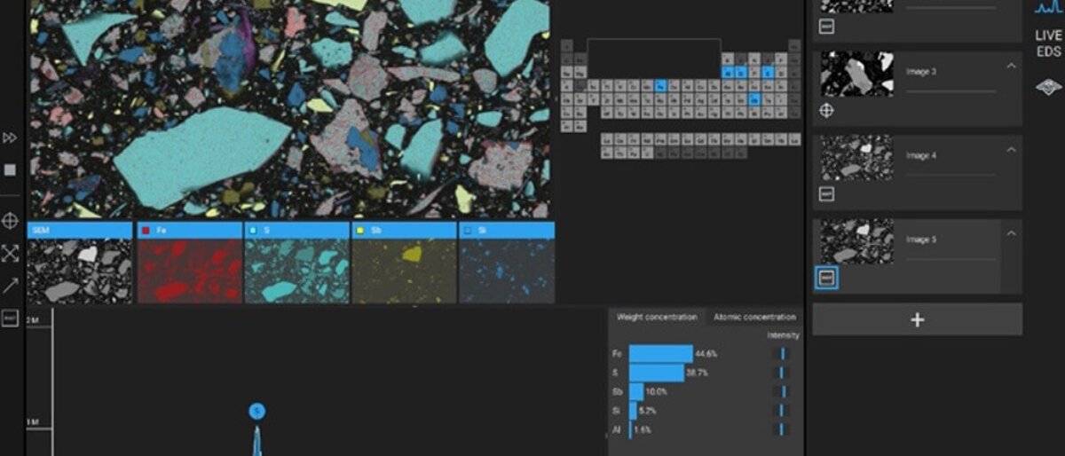
X-ray analytical mapping allows the operator to understand how composition varies over larger sample areas. Using the fast mapping option in the Phenom desktop SEM software, a colored display is accessible for larger areas. This feature shows initial results within seconds and refines and builds the image over time to achieve a reliable final result. By using the line-scan option, cross sections of one or more overlapping surfaces can also be analyzed.


Related posts
Download the full brochure on EDS in the desktop SEM here
- All
- Digital & Electronmicroscopy
- news

Tagarno T50 – A new digital microscope with 4K and 60 frames per second live images
Launching 2024, the TAGARNO T50 offers The best live image in the world with 4K and 60 frames per second images Automatic height adjustment with auto focus Low and high power magnification (up to 660x) on one single system Magnetic lens and ring light attachment Large working space User profiles and menu customization Software and [...]
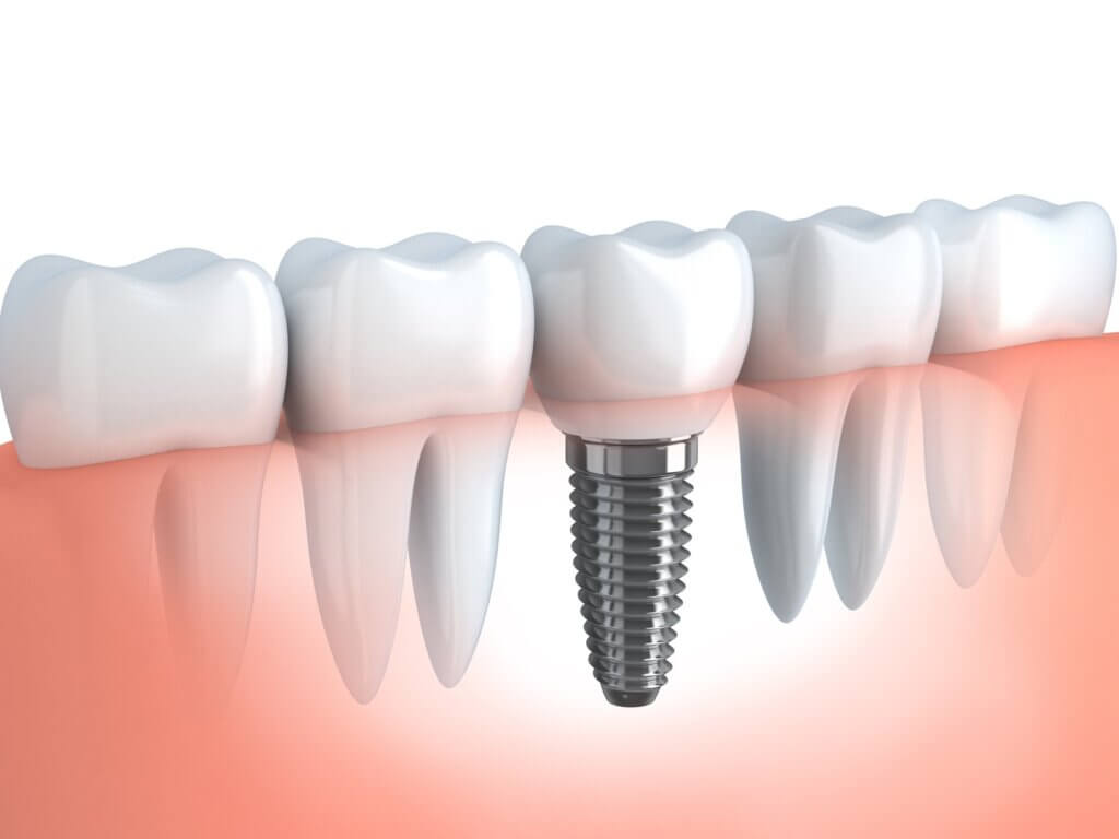
Desktop electron microscopy provides a quality seal for dental implants
In the world of dental implants, dentists and patients have to rely on manufacturer statements or FDA and CE marks to feel sure that the implants they use are being manufactured to a standard one would expect of an implantable dental device. However, recent studies of more than 250 dental implants from over 200 brands [...]

In need of steel-hard analyses?
Choose Phenom SEM
Steel is an infinitely recyclable and steel-hard material. Not surprisingly, it is used in a wide variety of structures: from bridges to vehicles to wind turbines. For just about every country in the world, steel is a crucial element in economic trade and production. A thorough analysis is recommended and Phenom SEM is the best [...]